Have you ever wondered what’s going on inside your head? Not just your thoughts, but the actual electrical activity racing through your brain? Welcome to the fascinating world of brainwaves and the science that allows us to peek into the mind’s inner workings: Electroencephalography, or EEG for short.
Imagine being able to see the invisible symphony of electrical pulses that make up your thoughts, emotions, and even your dreams. That’s exactly what Electroencephalography does. It’s like a stethoscope for your brain, letting scientists and doctors listen in on the constant chatter of your neurons. But why does this matter? From diagnosing epilepsy to unlocking the mysteries of consciousness, EEG has become an indispensable tool in understanding the most complex organ in the known universe – the human brain.
What is Electroencephalography (EEG)?
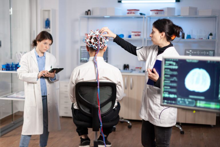
Electroencephalography (EEG) is a technique used to measure electrical activity in the brain by placing electrodes on the scalp. It helps researchers and doctors monitor brainwaves and diagnose neurological conditions like epilepsy.
What are Brainwaves?
Before we go into the technicalities of Electroencephalography EEG, let’s talk about what it’s actually measuring: brainwaves.
Brainwaves are rhythmic patterns of electrical activity produced by neurons in the brain. These waves are categorized into different types based on their frequency, each linked to specific mental states like relaxation, concentration, or deep sleep.
In other words, your brain is made up of billions of neurons, and these cells communicate with each other through tiny electrical pulses. When millions of neurons fire together, they create rhythmic patterns of electrical activity that we call brainwaves. Think of it like a crowd at a stadium doing “the wave” when enough people move together, you can see a pattern emerge.
Scientists have identified several types of brainwaves, each associated with different states of consciousness:
Delta waves (0.5-4 Hz): These slow waves dominate when you’re in a deep, dreamless sleep, like those nights when you wake up feeling completely rested, even if you don’t remember dreaming.
Theta waves (4-8 Hz): Often observed during light sleep, drowsiness, or deep meditation. Theta waves are linked to creative insights and the brain’s ability to visualize and daydream. Imagine the gentle, repetitive lapping of waves on a calm beach that’s the rhythm of your brain in this state.
Alpha waves (8-13 Hz): These appear when you’re relaxed but awake, like when you’re daydreaming or closing your eyes to rest. Alpha waves indicate that the brain is in a calm state, which alerts ‘idle’ mode, preparing for more focused activity.
Beta waves (13-30 Hz): These faster waves occur when you’re alert, focused, or problem solving. Think of the choppy waves on a windy day.
Gamma waves (30-100 Hz): The fastest waves, associated with peak concentration and potentially with moments of insight. These are like the rapid ripples caused by skipping a stone across water.
Understanding these different types of brainwaves helps researchers and clinicians interpret Electroencephalography readings and gain insights into a person’s mental state or potential neurological issues.
The History of Electroencephalography (EEG) and Brain Research
The story of Electroencephalography begins in 1875 when Richard Caton, a Liverpool physician, discovered electrical signals in animal brains. But it wasn’t until 1924 that Hans Berger, a German psychiatrist, recorded the first human EEG.
Berger’s interest in the brain was sparked after a near-death experience as a young man, which led him to believe in a form of telepathy. Driven by this curiosity, he eventually made his breakthrough in 1924, recording his son’s brain activity with crude radio equipment—a leap that seemed almost magical at the time. This primitive Electroencephalography was a far cry from today’s sophisticated machines, but it laid the groundwork for a revolution in neuroscience.
Over the decades, Electroencephalography (EEG) technology has evolved dramatically:
- 1930s-40s: Electroencephalography (EEG) gains recognition as a valuable diagnostic tool, particularly for epilepsy.
- 1950s: The development of Electroencephalography (EEG) topography allows for mapping brain activity across the scalp.
- 1970s-80s: Computerized Electroencephalography (EEG) analysis begins, allowing for more precise and complex data interpretation.
- 1990s-present: Advanced digital Electroencephalography (EEG) systems and new analysis techniques open up new research possibilities and clinical applications.
Today, Electroencephalography (EEG) continues to evolve, with new technologies like dry electrodes and portable devices making brain monitoring more accessible than ever before.
How Does EEG Work to Measure Brain Activity?
Now that we know what brainwaves are and how we discovered them, let’s go deeper into how modern EEGs work.
The Electroencephalography (EEG) is a straightforward powerful tool. It works by placing small, flat metal discs called electrodes on the scalp to measure the brain’s electrical activity. These electrodes are incredibly sensitive, detecting voltage changes as small as a few millionths of a volt when neurons communicate.
Step-by-Step Breakdown of the EEG Process:
Electrode Placement: The first step involves placing multiple electrodes on the scalp in a specific, standardized order. Typically, 16 to 25 electrodes are used in a clinical EEG, but in research settings, high-density EEGs can utilize up to 256 electrodes for more precise measurements. The positioning follows the “10-20 system,” which ensures consistent coverage of key areas of the brain. The purpose is to map out different sections of brain activity, giving a comprehensive view of how different parts of the brain are functioning.
Signal Detection: After the electrodes detect small voltage changes caused by the electrical firing of neurons. These changes are incredibly faint and often reflect the synchronized firing of groups of neurons. While a single neuron firing is undetectable, the collective activity of many neurons generates enough electrical energy for the electrodes to pick up on. The signals captured are not from deep within the brain but primarily from the surface areas like the cerebral cortex. These signals give us insight into overall brain function and activity patterns.
Amplification: Since the electrical signals from the brain are extremely weak, they must be amplified. The EEG machine boosts these signals thousands of times to make them strong enough for analysis. Without this step, the brain’s electrical activity would be too subtle to observe. The amplified signals allow us to “hear” and analyze what’s happening within the brain, turning otherwise invisible brain activity into readable data.
Recording: The next stage is recording these amplified signals. Historically, this was done on moving paper, with ink tracing the wave patterns in real time. Today, EEG signals are more often recorded digitally on a computer, making it easier to store, analyze, and share data. This change to digital storage also opens the way to more advanced software tools that can provide faster and more accurate interpretations of the data collection.
Analysis: After recording is done, the real work begins. The raw data captured by the EEG is complex and needs to be processed and interpreted. Specialized software is often used to break down the signals into different types of brainwaves which are: alpha, beta, delta, and theta waves. Each type of wave is associated with different mental states. For example, alpha waves are often linked to relaxed, calm states, while beta waves are more common during active thinking or concentration. Analyzing these brainwave patterns can help doctors diagnose neurological conditions or monitor cognitive states.
It’s important to note what EEG can and can’t tell us. EEG is excellent at showing the overall rhythms and patterns of brain activity, making it invaluable for diagnosing conditions like epilepsy or sleep disorders. It also has unparalleled temporal resolution, meaning it can track changes in brain activity down to the millisecond.
However, EEG has limitations. It primarily detects activity near the surface of the brain, so it can’t give us detailed information about deeper brain structures. It also can’t tell us exactly which neurons are firing or why. For these kinds of insights, we need to combine EEG with other brain imaging techniques.
Applications of Electroencephalography (EEG)
The applications of EEG are vast and continually expanding. Here are some of the key areas where EEG makes a significant impact:
Medical Uses:
Epilepsy diagnosis and management: EEG can detect the abnormal brain activity that causes seizures.
Sleep disorders: EEG is an important component of sleep studies, helping diagnose conditions like narcolepsy or sleep apnea.
Brain injuries: EEG can help assess the extent of brain damage and monitor recovery.
Coma and brain death: EEG is used to evaluate brain activity in unresponsive patients.
Research Applications:
Cognitive science: EEG helps researchers study attention, memory, and other cognitive processes.
Psychology: It’s used to investigate emotional responses, decision-making, and mental health conditions.
Linguistics: EEG can provide insights into language processing and acquisition.
Emerging Fields:
Brain-Computer Interfaces (BCIs): EEG signals can be used to control external devices, offering real hope for paralyzed individuals.
Neurofeedback: People can learn to modify their brain activity by watching real-time EEG readouts.
Consumer applications: Simple EEG devices are now available for meditation assistance, sleep tracking, and even gaming control.
Limitations and Challenges of Electroencephalography (EEG)
While EEG is a powerful tool, it’s not without its limitations and challenges:
Common Misconceptions:
EEG doesn’t read thoughts: It measures brain activity, not specific thoughts or memories. – It’s not a lie detector: While EEG can show stress responses, it can’t definitively detect deception.
Technical Challenges:
Signal noise: EEG is sensitive to electrical interference from muscle movements, eye blinks, and even nearby electronic devices.
Spatial resolution:EEG has poor spatial resolution compared to techniques like fMRI, which has led to debates in the scientific community about the accuracy of its interpretations. However, despite these debates, its unmatched temporal resolution keeps EEG at the forefront of real-time brain activity monitoring.
Interpretation Difficulties:
Complexity: EEG data is complex and can be challenging to interpret, requiring specialized training.
Individual differences: Brain activity patterns can vary significantly between individuals, making standardization difficult.
The Future of Electroencephalography (EEG)
Despite these challenges, the future of EEG looks bright. Emerging technologies and methodologies are expanding its capabilities:
High-density EEG systems, which can have hundreds of electrodes (quite the number, right?), are showing promise in improving spatial resolution.
Dry electrodes: These could make EEG setup faster and more comfortable, enabling long-term monitoring.
AI and machine learning: Advanced algorithms are improving EEG data analysis and interpretation.
Combining EEG with other techniques: Integrating EEG with fMRI or MEG provides more comprehensive brain imaging.
New innovations are on the horizon. For example, startups are already developing consumer-friendly EEG devices for mental health tracking, while partnerships between tech companies and research institutions could soon offer early detection of Alzheimer’s or personalized treatments for ADHD..
Advanced BCIs that could restore communication or movement to severely paralyzed individuals.
Personalized medicine approaches using EEG to predict individual responses to psychiatric medications.
Conclusion
From its humble beginnings in the early 20th century to today’s cutting-edge applications, EEG has revolutionized our understanding of the brain. It allows us to peer into the electrical symphony that underlies all our thoughts, feelings, and actions.
As we’ve explored, EEG is more than just a medical diagnostic tool. It’s a window into the very essence of what makes us human, our consciousness. Whether it’s helping doctors treat epilepsy, enabling researchers to unravel the mysteries of sleep, or empowering paralyzed individuals to communicate, EEG continues to push the boundaries of neuroscience.
As technology advances, who knows what new insights and applications we’ll discover? One thing is certain: the story of EEG and our exploration of brainwaves is far from over. So, the next time your mind starts to wander, think about the hidden symphony of brainwaves orchestrating your every thought. If you’re curious to go deeper, consider exploring real-time brainwave tracking apps or reading up on how EEG is influencing fields like cognitive enhancement. The world of EEG is growing, and who knows your next insight could come from your own brain. Don’t forget to leave a comment and share this post for others to understand where we’re heading.

